Features
Finance
Management
The Feet & Their Slings
The foot is our point of contact with the ground. Uniquely, the human species transfers all the weight of our long bodies down though the delicate collection of bones that is the foot.
September 9, 2009 By Mark Finch KMI
The foot is our point of contact with the ground. Uniquely, the human species transfers all the weight of our long bodies down though the delicate collection of bones that is the foot.
Looking at the bones one would think this delicate collection of unstable bones is no candidate for such an important job.
Not only does the foot need to provide support but also absorb shock, transfer force and provide the spring in our step. The bones clearly need some help.
A closer look at the shape of the bones reveals a series of arches and wedges that begin to provide stability, but clearly this is not enough.
The foot gains stability and strength from the tough ligaments that surround the bones and from the pull of the muscles coming down from the lower leg. These muscles descend from the leg and wrap under the foot to create a kind of a stirrup or sling.
The foot is supported by three arches; the medial, transverse and longitudinal. The longitudinal arch runs along the outside of the foot on the little toe side.
There may not seem to be much of an arch on the outside of your foot, but under the fleshy tissue there is a boney arch that supports the outer edge of the foot.
The transverse arch, as the name suggests runs across the foot from the big toe out to the little toe. This arch is well supported with tough connective tissue.
 |
|
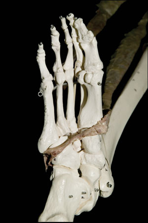
| Figure 1 A cadaver dissection showing the continuity of the tibialis anterior and peroneus longus muscles along the bottom of the foot. The dissected tissue has been laid on a plastic skeleton to show the geography. Anatomy Trains , 2nd ed. Elsevier. |
The medial arch is the largest and most significant of the three.
It runs from the heel to the big toe. It is involved in the springing off of the foot and is required to be flexible to allow the full range of foot movements.
The medial arch is the most mobile of the arches but also the least stable. More than the other arches, this one relies on myofascial (muscle and fascia) support to maintain its shape.
The largest and most significant myofascial support for the foot is the plantar fascia (or plantar aponneurosis). This tough piece of fascia forms a spring under the foot, which supports all the arches in weight bearing. The plantar fascia cannot be considered a tendon, as it’s not firmly attached to the heel bone. It can however be seen as a continuation of the calf muscles.
 |
|
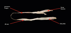
| Figure 2 (Above): A cadaver dissection of the lower (or leg) portion of the Spiral Line. It shows that the continuity doesn’t stop with the peroneus longus and tibialis anterior but continues to include the biceps femoris and illiotibial tract (including the tensor fascia lata), thus joining the front of the pelvis to the back of the pelvis by way of the arch of the foot. Anatomy Trains , 2nd ed. Elsevier. • Both photos used with kind permission by Thomas Myers. • Thomas Myers, Anatomy Trains Revealed, Early Dissective Evidence. Available on DVD at: www.AnatomyTrains.com |
In the Anatomy Trains model (Myers 2001) myofascial continuums are described as crossing bones and joints. In this way we can see that the calf muscles of gastrocnemius and soleus blend into the achillies tendon, which is continuous with heel fascia and then the plantar fascia. In this way the plantar fascia can be considered part of a myofascial continuum that supports all the arches from collapsing under the weight of the body. So although not a tendon the plantar fascia is the most supporting piece of myofascia for the arches.
Now let’s explore the tendons proper that provide support for the arches. There are five muscles in total that directly contribute to the support of the arches by pulling up from above.
Three of these muscles form two stirrups or slings. The first and most significant myofascial sling comes from the frequently documented connection between the tibialis anterior and peroneus longus.
Starting at the head of the fibula, the peroneus longus slides straight down the lateral leg, behind the medial malleolus, and under the foot to the base of the medial cuneiform (and the base of the first metatarsal).
From the medial cuneiform there is a continuous connection with the tendon of the tibialis anterior (figures 1 and 2), which sweeps back across the ankle up to the lateral tibia.
Together, these myofascial units form a sling that controls the movement of the medial arch. Let’s call this the sling of the lower leg. It is most active and useful in simple standing when both sets of muscle are in the middle of their range and therefore have the biggest mechanical advantage.
To feel this sling, try standing on one leg, can you feel the front of the shin working and the small movements in the medial arch? This is the lower leg sling adjusting to the load through one leg and making small compensatory movements to keep the medial arch supported.
If this is hard to feel, just stay in this position and wait for the muscles to fatigue, or close your eyes. Then you should be able to feel the lower leg sling working to stabilize the medial arch in standing.
What happens though when the ankle is not in the middle of the range and is in plantar flexion? Imagine you are on your tiptoes getting a jar out of that very top cupboard, or a ballerina on ‘pointe.’
Here, the tibialis anterior is in a lengthened position and at the end of its range so has a limited mechanical advantage and can provide limited stabilization for the medial arch. The body solves this problem by switching part of the sling to a deeper aspect of the leg. The peroneus longus remains the outside part of the sling and from the medial cuneiform the sling continues to the posterior aspect of the leg with the tibialis posterior.
This muscle is also continuous with the peroneus longus and instead of switching back across the ankle to the front of the tibia; it tucks behind the ankle and finds its attachment on the backside of the tibia.
Let’s call this the deep lower leg sling. When the foot is pointed these muscles are in the middle of their range and therefore have a good possibility of stabilizing the medial arch when the heel is raised off the ground. So the deep lower leg sling stabilizes the medial arch when the heel is off the ground.
Print this page